Home

capacità Tuttavia labirinto collagen tem pipistrello insegnante Uomo

False-colour Tem Of Collagen Fibres Photograph by J Gross, Biozentrum |  Fine Art America
False-colour Tem Of Collagen Fibres Photograph by J Gross, Biozentrum | Fine Art America

Application of Detergents or High Hydrostatic Pressure as Decellularization  Processes in Uterine Tissues and Their Subsequent Effects on In Vivo  Uterine Regeneration in Murine Models | PLOS ONE
Application of Detergents or High Hydrostatic Pressure as Decellularization Processes in Uterine Tissues and Their Subsequent Effects on In Vivo Uterine Regeneration in Murine Models | PLOS ONE

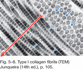
File:Fibers of Collagen Type I - TEM .jpg - Wikimedia Commons

Cryo-TEM Analysis of Collagen Fibrillar Structure - ScienceDirect

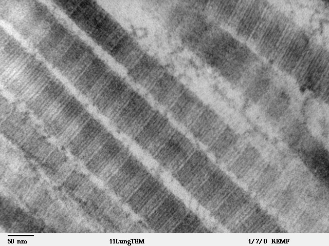
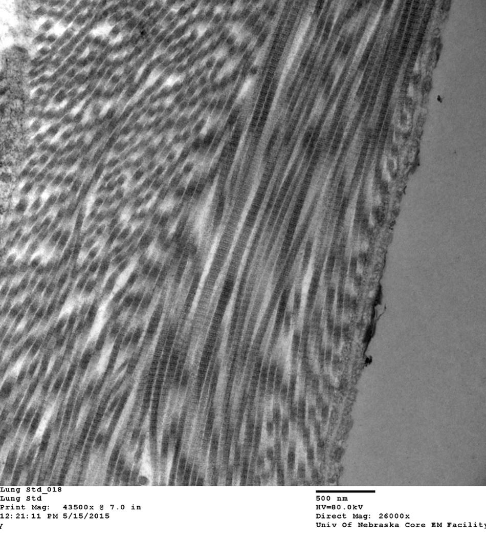
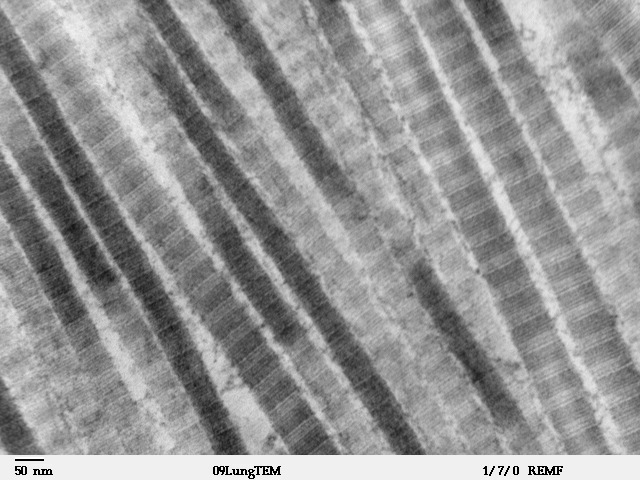
Collagen Fibrils in Lung Tissue | VCR | University of Nebraska Medical  Center
Collagen Fibrils in Lung Tissue | VCR | University of Nebraska Medical Center

Human Structure Virtual Microscopy

Collagen damage - TEM | Wellcome Collection

Electron Microscope Observation of Collagen fibers

Transmission electron micrograph (TEM) showing the typical corneal stroma  or substantia propria. It is composed of layers of collagen fibres, forming  Stock Photo - Alamy
Transmission electron micrograph (TEM) showing the typical corneal stroma or substantia propria. It is composed of layers of collagen fibres, forming Stock Photo - Alamy

Poisson's ratio of collagen fibrils measured by small angle X-ray  scattering of strained bovine pericardium: Journal of Applied Physics: Vol  117, No 4
Poisson's ratio of collagen fibrils measured by small angle X-ray scattering of strained bovine pericardium: Journal of Applied Physics: Vol 117, No 4

File:Fibers of Collagen Type I - TEM.jpg - Wikimedia Commons

Type III Collagen Can Be Present on Banded Collagen Fibrils Regardless of  Fibril Diameter
Type III Collagen Can Be Present on Banded Collagen Fibrils Regardless of Fibril Diameter

Transmission electron microscopy (TEM) images of collagen structure... |  Download Scientific Diagram
Transmission electron microscopy (TEM) images of collagen structure... | Download Scientific Diagram

Science Source Stock Photo - Collagen Fibers (TEM)

Collagen Fibres, Tem Photograph by Thomas Deerinck, Ncmir | Pixels

Porcine cornea stromal collagen architechture, TEM, Transmission electron  microscopy | Animal print rug, Printed rugs, Animal print
Porcine cornea stromal collagen architechture, TEM, Transmission electron microscopy | Animal print rug, Printed rugs, Animal print

Non-Enzymatic Decomposition of Collagen Fibers by a Biglycan Antibody and a  Plausible Mechanism for Rheumatoid Arthritis | PLOS ONE
Non-Enzymatic Decomposition of Collagen Fibers by a Biglycan Antibody and a Plausible Mechanism for Rheumatoid Arthritis | PLOS ONE

Connective Tissue Quiz Answer - Slide #7

Connective Tissue - Slide #21

Transmission electron microscopy (TEM) shows evidence of highly ordered...  | Download Scientific Diagram
Transmission electron microscopy (TEM) shows evidence of highly ordered... | Download Scientific Diagram

Optical Microscopy and Electron Microscopy for the Morphological Evaluation  of Tendons: A Mini Review - Xu - 2020 - Orthopaedic Surgery - Wiley Online  Library
Optical Microscopy and Electron Microscopy for the Morphological Evaluation of Tendons: A Mini Review - Xu - 2020 - Orthopaedic Surgery - Wiley Online Library

Morphological Characteristics of the Dermal Papillae in the Development of  Pressure Sores
Morphological Characteristics of the Dermal Papillae in the Development of Pressure Sores

Cryo-TEM images of a collagen fiber during biomineralization. (a) At a... |  Download Scientific Diagram
Cryo-TEM images of a collagen fiber during biomineralization. (a) At a... | Download Scientific Diagram

Collagen fibres, TEM - Stock Image - C036/7437 - Science Photo Library

Using transmission electron microscopy and 3View to determine collagen  fibril size and three-dimensional organization | Nature Protocols
Using transmission electron microscopy and 3View to determine collagen fibril size and three-dimensional organization | Nature Protocols
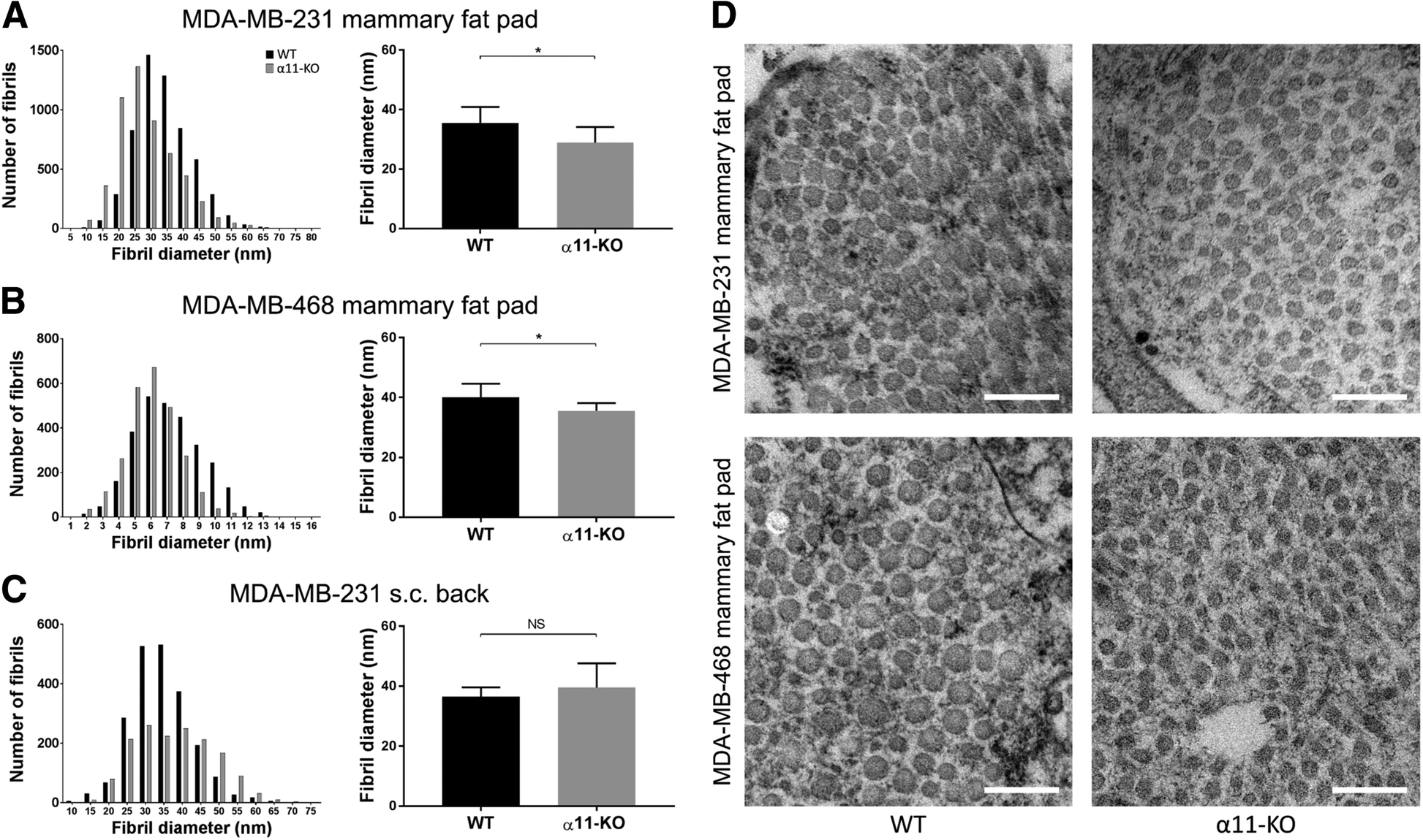
Stromal integrin α11-deficiency reduces interstitial fluid pressure and  perturbs collagen structure in triple-negative breast xenograft tumors |  BMC Cancer | Full Text
Stromal integrin α11-deficiency reduces interstitial fluid pressure and perturbs collagen structure in triple-negative breast xenograft tumors | BMC Cancer | Full Text

Collagen Fibril Diameter by TEM.

Structural changes in collagen fibrils across a mineralized interface  revealed by cryo-TEM - ScienceDirect
Structural changes in collagen fibrils across a mineralized interface revealed by cryo-TEM - ScienceDirect

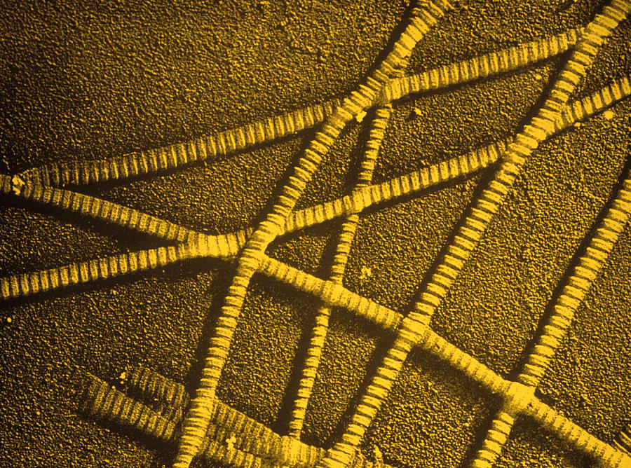
TEM results of collagen mineralization after almost 1 week of reaction....  | Download Scientific Diagram
TEM results of collagen mineralization after almost 1 week of reaction.... | Download Scientific Diagram